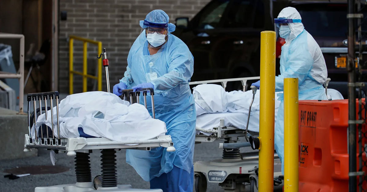
Pandemic threat? Anyone else concerned?
- Thread starter erkme73
- Start date
You are using an out of date browser. It may not display this or other websites correctly.
You should upgrade or use an alternative browser.
You should upgrade or use an alternative browser.
mat200
IPCT Contributor
- Jan 17, 2017
- 16,479
- 27,688
So this is partly true, it was not created in China, it was created in the U.S. then sent to China to test...
Another partly true statement is the last sentence, yes it was transmitted to humans, to Chinese humans, from another species, yes under the direction of Fauci and his Evil Scientist (from another species) wanting to KILL Humans...

Glad to see NPR defunded for spreading Misinformation...Thank You Trump...
bigredfish
Known around here
bigredfish
Known around here
Oceanslider
Known around here
bigredfish
Known around here
bigredfish
Known around here
Very well explained and informative...
My son and his wife decided to not vaccinate their son, who is now 8 months old and has Never been sick, not even a cold even though both of their parents have had colds/sinuses a few times in this 8 months. Their choice, I have been on the fence on this only because we grew up trusting Doctors, but today after going through the Plandemic and learning so, so much of what really went on these past decades I am almost completely off the fence towards no vaccines, I know my wife and I are done with any future vaccines with all the push of mRNA vaccines. Time will tell but I will share a story told to us by our Tree guy about two children in his family, both around the same age, both with parents who are related, think he said brother/sister (No they are not married to each other, lol, yea they live out in the country, haha) . One is not vaccinated, the other is...The unvaccinated is an extremely healthy child, never sick yet is around other kids like the other child who is vaccinated and is Always sick. This is not the first time I have heard this. Yes I know, genetics needs to be looked at along with many other things, their parents and so on. But the core of this is unvaccinated people with a healthy diet/immune system/lifestyle seem to be less likely to have a compromised immune system.
The fact that we have a law that makes all vaccine companies immune from being sued is a HUGE RED FLAG...
Would you buy a car or anything from a company that has Zero responsible if their product is defective? Of course not, yet we do with our health? Wow, how brainwashed are we?
My son faces not being able to take his son to daycares, schools, etc. due to their requirement that the child is vaccinated? Do you see the irony in this. Forced Vaccination. Shun the unvaccinated from society? This tells me that the 29 vaccines our present day children get Don't Work...
Many, many, many, many, many questions I have...
bigredfish
Known around here
Young couples with children like you describe are facing a huge dilemma with lots of hard choices. I feel for them
bigredfish
Known around here
bigredfish
Known around here

Insurance Industry Data Exposes '5,000 Vaccine-Linked Deaths a WEEK' - Slay News
One of the world's leading data experts has revealed that the insurance industry is now seeing up to 5,000 deaths every single week that are linked to Covid mRNA "vaccines."
 slaynews.com
bigredfish
Known around here

Insurance Industry Data Exposes '5,000 Vaccine-Linked Deaths a WEEK' - Slay News
One of the world's leading data experts has revealed that the insurance industry is now seeing up to 5,000 deaths every single week that are linked to Covid mRNA "vaccines."slaynews.com
And yet no public outcry.
Those who got jabbed can’t bring themselves to admit it. Those who didn’t get jabbed can’t say “told you so” as it’s not polite, and government can’t admit it because the repercussions would be too great
bigredfish
Known around here
There just can't be enough reports to get our wonderful gov't to stop this madness can there?

 slaynews.com

Scientists Sound Alarm as Deadly Abnormalities Found in mRNA 'Boosted' Healthy Young Adults - Slay News
A group of scientists has just issued a chilling warning after a major new study confirmed that dangerous abnormalities are triggering in healthy young adults within hours of receiving a Covid mRNA "booster" injection.
 slaynews.com
bigredfish
Known around here
And here's the actual study.
 www.frontiersin.org
Its truly amazing that:
1- these kill shots are still being recommended by our FDA/CDC because the fucking fraud/Grifter-in-Chief can't admit he was wrong
2- a segment of the population hear and read these type of reports, and continue doing as they're told because they've been so programmed by their government and institutions and have lost the ability to think for themselves
Honestly I'm past giving a shit about the Jab enthusiasts, let 'em die if they are so intent on doing so. It will help cleanse the Gene pool
Frontiers | Immune and hematologicak responses to the third dose of an mRNA COVID-19 vaccine: a six-month longitudinal study
The deployment of mRNA vaccines against SARS-CoV-2 is a major landmark in controlling the COVID-19 pandemic. However, the activation of adaptive immunity and...
Its truly amazing that:
1- these kill shots are still being recommended by our FDA/CDC because the fucking fraud/Grifter-in-Chief can't admit he was wrong
2- a segment of the population hear and read these type of reports, and continue doing as they're told because they've been so programmed by their government and institutions and have lost the ability to think for themselves
Honestly I'm past giving a shit about the Jab enthusiasts, let 'em die if they are so intent on doing so. It will help cleanse the Gene pool
Last edited:

